Packs sizes (2)
| code | packaging size | price per unit | box price per unit | |
|---|---|---|---|---|
| Code & packaging | Price per piece | |||

|
code
251338.1610
|
packaging size
500 ml
|
price per unit
single
88,20€
|
box price per unit
74,97€x 6 units
|

|
code
251338.1211
|
packaging size
1000 ml
|
price per unit
single
122,50€
|
box price per unit
104,13€x 6 units
|
Technical data
- Density:
- 1.058 kg/l
- Physical Description:
- liquid
Blue with red shades liquid
- Product Code:
- 251338
- Product Name:
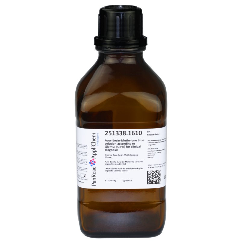
- Giemsa's Azur-Eosin-Methylene Blue solution(slow)(CE-IVD) for clinical diagnostics
- Quality Name:
- for clinical diagnostics
- Headline Comment:
- for hematology, blood smear staining
- Specifications:
- COMPOSITION:
Azur-Eosin-Methylene Blue dye according to Giemsa: 0.5 g
Methanol: 50 ml
Glycerol: 50 ml
Maximum limit of impurities
Suitability for hematology stain: passes test
- Hazard pictograms
-
- UN:
- 1230
- Class/PG:
- 3(6.1)/II
- ADR:
- 3(6.1)/II
- IMDG:
- 3(6.1)/II
- IATA:
- 3(6.1)/II
- WGK:
- 1
- Storage:
- Room Temperature.
- Signal Word:
- Danger
- GHS Symbols:
- GHS02
GHS06
GHS08
- H Phrases:
- H225
H331
H311
H301
H370
- P Phrases:
- P210
P233
P240
P241
P242
P501
P243
P260
P261
P264
P270
P271
P280
P301+P310
P302+P352
P303+P361+P353
P304+P340
P307+P311
P311
P312
P321
P322
P330
P361
P363
P370+P378
P403+P233
P403+P235
P405
- Master Name:
- Giemsa's Azur-Eosin-Methylene Blue solution (slow)
- CS:
- 38229000
Documents
Inquiry
Comments
HistologyHistology is the area of biology that studies the composition, structure, and characteristics of the organic tissues of living beings. Histology is closely related to microscopic anatomy, as its study does not stop at tissues, but goes beyond them, also observing cells internally and other corpuscles, and is related to biochemistry, cytology and medicine. - Histology has become very important in medicine and biology as it is crucial for these disciplines because it’s in between the intersections biochemistry, molecular biology, and physiology on the one hand and pathological processes and their consequences on the other. - Classical histology techniques are applied in medical laboratories for the diagnosis of a wide variety of diseases. These techniques are adequate in most diagnostic cases. However, when diagnosis with these classical techniques cannot be considered reliable, additional methods must be used. These techniques include histochemical staining, immunohistochemical methods, DNA hybridization, fluorescence in situ hybridization (FISH), PCR, flow cytometry and others. - The medical laboratories mentioned above focus on primarily on production-based applied science (number of diagnostics tests), as opposed to research laboratories which focus on basic science on an academic basis. - A medical laboratory or clinical laboratory is the place where tests are usually performed on clinical samples to obtain information about a patient's health in terms of diagnosis, treatment, and prevention of disease. - Research laboratories use conventional techniques for Genomics, Proteomics and Cell Culture procedures. - PanReac AppliChem branded products for hospital laboratories: - Medical laboratories: Microscopy products. - Research Laboratories: Genomics, Proteomics and Cell Culture Products. - Medical laboratories - In many countries there are mainly two types of medical laboratories depending on the type of research performed. - Hospital laboratories: laboratories integrated into a hospital to perform tests on patients’ samples. There are 4 different types of laboratories: - Clinical pathology laboratories: hematology, histopathology, cytology, routine pathology. - Clinical microbiology laboratories: bacteriology, mycobacteriology, virology, mycology, parasitology, immunology, serology. - Clinical biochemistry laboratories: biochemical analyses, hormone assays, etc. - Molecular diagnostic laboratory or cytogenetics and molecular biology laboratory. - External clinical laboratories: for extremely specialized tests, the sample can go to an external research laboratory. - ITW Reagents offers a complete range of products for histology, hematology, and microbiology under the PanReac AppliChem brand, including the most common used reagents in the process of sample preparation for microscopic examination. This range covers all stages of fixation, clearing, paraffin embedding, staining and mounting. ITW reagents also have a wide range of products for research in different areas in Life Sciences for assays to be developed in hospital laboratories: genomics, proteomics, and cell culture. - Many of the PanReac AppliChem branded products used in microscopy carry the CE IVD marking in compliance with the new European Regulation (EU) 2017/746 on in vitro diagnostic products (IVDR). - Histological processing techniques and stages - Although most of the stages of histological processing are common, some of them are unique to a single type of sample processing. For example, embedding is done only on tissues and heat fixation only on blood samples. - The most common stages and the products used are as follows: - Fixation: Fixation interrupts degradation processes that occur after cell death, trying to preserve the tissue/cellular architecture and composition as close as possible to how it was in the living organism. The most common type is chemical fixation with Formaldehyde 3.7-4.0% w/v buffered to pH=7 and stabilized with methanol and, for increased safety, Histofix® Formaldehyde single-dose ready-to-use preservative. - Dehydration and clearing: Dehydration is the complete removal of water from the specimen or tissue sample so that it can be properly embedded in the subsequent non-water-soluble embedding media. The fixed and washed specimens are passed through 96% alcohol and then absolute alcohol for varying lengths of time, typically one and a half hours in each bath. Clearing is the replacement of the drying agent with a substance miscible with the embedding medium to be used. The most common products in this stage are alcohols such as absolute ethanol, 96% ethanol, 70% ethanol, which can be denatured or not, as well as xylene. - Inclusion: by inclusion, the water in the tissue is replaced by a liquid medium capable of solidifying under suitable temperature conditions. Paraffin wax P.F. 55-58°C plasticized + DMSO in lentils or simply Paraffin wax P.F. 56-58°C in lentils is used for this purpose. - Cutting: Paraffin-embedded tissues are cut thinly enough (4-6 microns) to allow light to pass through for examination under a microscope. This is done with a microtome: a mechanical instrument used to make micrometric sections of tissue. We recommend our paraffin cleaner to keep the microtome clean. - Clearing-Hydration: Dewaxing-hydration is the process of removing the embedding medium from paraffin-embedded tissue sections and rehydrating them for proper dye penetration. The most common reagents at this stage are alcohols such as Absolute Ethanol, Ethanol 96%, Ethanol 70% which may or may not be denatured in addition to Xylene. - Staining: Most tissues, especially those of animal origin, are colorless unless they contain pigment. It is therefore necessary to stain them for microscopic observation. This is achieved using dyes, substances which, in contact with a suitable support, bind to it in a durable way, transmitting its color. The most used dyes are hematoxylin and eosin, but there is a wide variety of stains depending on the tissue to be stained. - Mounting and immersion: Once the preparations have been rinsed, they must be mounted definitively. Mounting agents can be aqueous or non-aqueous; the type used depends on the relevant protocol. Examples of mounting media are DPX, Canada Balsam and Eukitt®. Immersion media are liquids, often natural oils, which have a defined refractive index. It is important that the refractive index is approximately 1.5 (same index as glass). This allows for a homogeneous oil immersion. For this purpose, Immersion Oil or Cedar Oil is used. - Microscopic analysis: This is the microscopic observation phase. No additional reagents are needed.


